
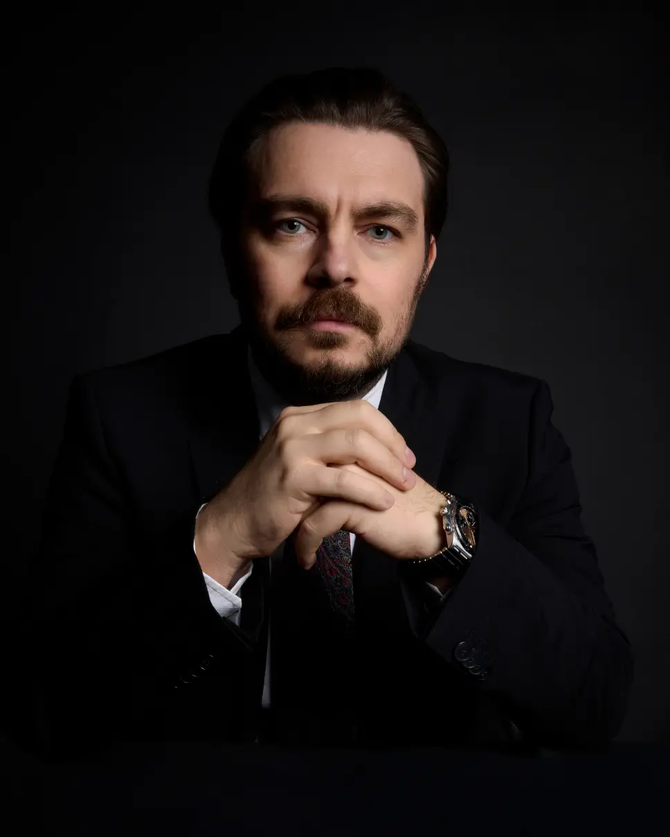
This article was written by Assoc. Prof. Dr. Burak Sercan Erçin and is based on clinical experience. A specialist in Plastic, Reconstructive and Aesthetic Surgery; prepared in accordance with current medical literature and personal surgical data. A consultation is recommended before making any medical decisions.
What is an Orbital Box Osteotomy?
It is the correction of severe orbital hypertelorism that represents a complicated of craniofacial surgery. Unlike routine aesthetic procedures that address the soft tissue envelope or superficial osseous contours, the Orbital Box Osteotomy (OBO) involves the fundamental dismantling and reconfiguration of the craniofacial skeleton. It is a procedure of immense complexity, requiring a convergence of neurosurgical access, ophthalmological preservation, and reconstructive artistry. The primary objective is the medial translocation of the bony orbits, essentially detaching the eye sockets from the skull base and moving them closer together to correct an abnormal interorbital distance.
Historically confined to a select few academic institutions in France, the United States, and the United Kingdom, the landscape of high-acuity craniofacial surgery is undergoing a significant geopolitical shift. The rising costs of healthcare in the West, coupled with the globalization of medical expertise, has positioned nations like Turkey as pivotal hubs for complex reconstruction. This guide provides an exhaustive analysis of the Orbital Box Osteotomy, ranging from its embryological origins and anatomical foundations to the granular details of surgical execution. Furthermore, it examines the logistical and clinical framework required for international patients to access this care, highlighting the emerging prominence of reconstructive specialists such as Associate Professor Dr. Burak Sercan Ercin in Istanbul.
The scope of this blog extends beyond a mere procedural description. It serves as a critical resource for understanding the physiological risks, the necessity of microsurgical contingency planning, and the intricate architecture of postoperative recovery. By synthesizing data on surgical outcomes, cost-benefit analyses, and international patient protocols, this guide aims to provide a definitive resource for stakeholders navigating the complex domain of craniofacial medical tourism.
Click the link above to watch the highlight
Anatomical and Pathophysiological Foundations
To comprehend the magnitude of an Orbital Box Osteotomy, one must first understand the pathology it aims to correct. Orbital hypertelorism is not a syndrome in itself but a physical manifestation—a sign—of an underlying craniofacial dysmorphology.
The Embryology of the Midface
The human face develops from five primordia that surround the stomodeum (primitive mouth): the single frontonasal prominence and the paired maxillary and mandibular prominences. During the early weeks of gestation (weeks 4-8), the primitive eyes are positioned laterally on the embryo’s head, similar to a fish or bird. As the frontonasal prominence differentiates and the maxillary processes grow medially, the orbits rotate forward and migrate toward the midline. This migration is essential for the development of overlapping visual fields, which confers stereopsis (depth perception).
Orbital hypertelorism results from a disruption in this morphogenetic movement. Several mechanisms have been proposed. First is premature ossification, where early fusion of the lesser wings of the sphenoid bone can physically lock the orbits in a lateral position, preventing medial migration. Second is physical obstruction; the presence of a facial cleft or an anterior encephalocele (a herniation of brain tissue through a skull defect) can act as a physical wedge, forcing the orbits apart. The “morphogenetic capsule” of the nasal cavity may fail to narrow, maintaining a widened interorbital distance. Finally, in syndromes such as Apert or Crouzon, the premature fusion of cranial sutures (coronal, sagittal, or lambdoid) alters the growth vectors of the skull base, often resulting in a shallow, wide orbital configuration.
Metric Classification and Diagnosis
The diagnosis of hypertelorism is quantitative. While “wide-set eyes” can be a subjective aesthetic trait (telecanthus), true orbital hypertelorism is defined by the underlying skeletal distance between the dacryons (the junction of the lacrimal, maxillary, and frontal bones) or the interorbital distance (IOD) measured on computed tomography (CT) scans.
In a healthy adult population, the mean interorbital distance is approximately 25 mm for females and 28 mm for males. Paul Tessier, the father of craniofacial surgery, established the severity grading that remains in use today. Degree I (Mild) is defined by an interorbital distance of 30 to 34 mm. These cases are often aesthetic but may not require transcranial surgery. Degree II (Moderate) involves an interorbital distance of 34 to 40 mm, where functional impairments begin to manifest. Degree III (Severe) is characterized by an interorbital distance greater than 40 mm. In extreme cases, distances exceeding 50 mm have been recorded. This level almost invariably requires an Orbital Box Osteotomy or facial bipartition for correction.
The Functional Sequelae of Hypertelorism
The implications of uncorrected severe hypertelorism extend far beyond the cosmetic stigma. The lateralization of the orbits has profound effects on the visual and respiratory systems.
As the orbits rotate outward, the visual axes diverge, leading to exotropia (wall-eyed appearance). The brain, unable to fuse the two disparate images, may suppress one eye to avoid diplopia (double vision), leading to amblyopia (lazy eye) and a permanent loss of binocular vision. Frequently, the deformity is not just horizontal but vertical. One orbit may be positioned lower than the other (vertical dystopia), requiring the surgical correction to include a vertical vector of movement.
In syndromic cases, such as Crouzon syndrome, the orbits are not only wide but shallow (exorbitism). The globe of the eye protrudes forward (proptosis), preventing the eyelids from closing completely during sleep (lagophthalmos). This chronic exposure can lead to drying of the cornea, ulceration, scarring, and eventually blindness. The Orbital Box Osteotomy, therefore, is a functional salvage procedure. By deepening the orbits and aligning the visual axes, the surgeon aims to protect the cornea and restore the potential for binocular vision, while simultaneously reconstructing a normalized facial appearance.
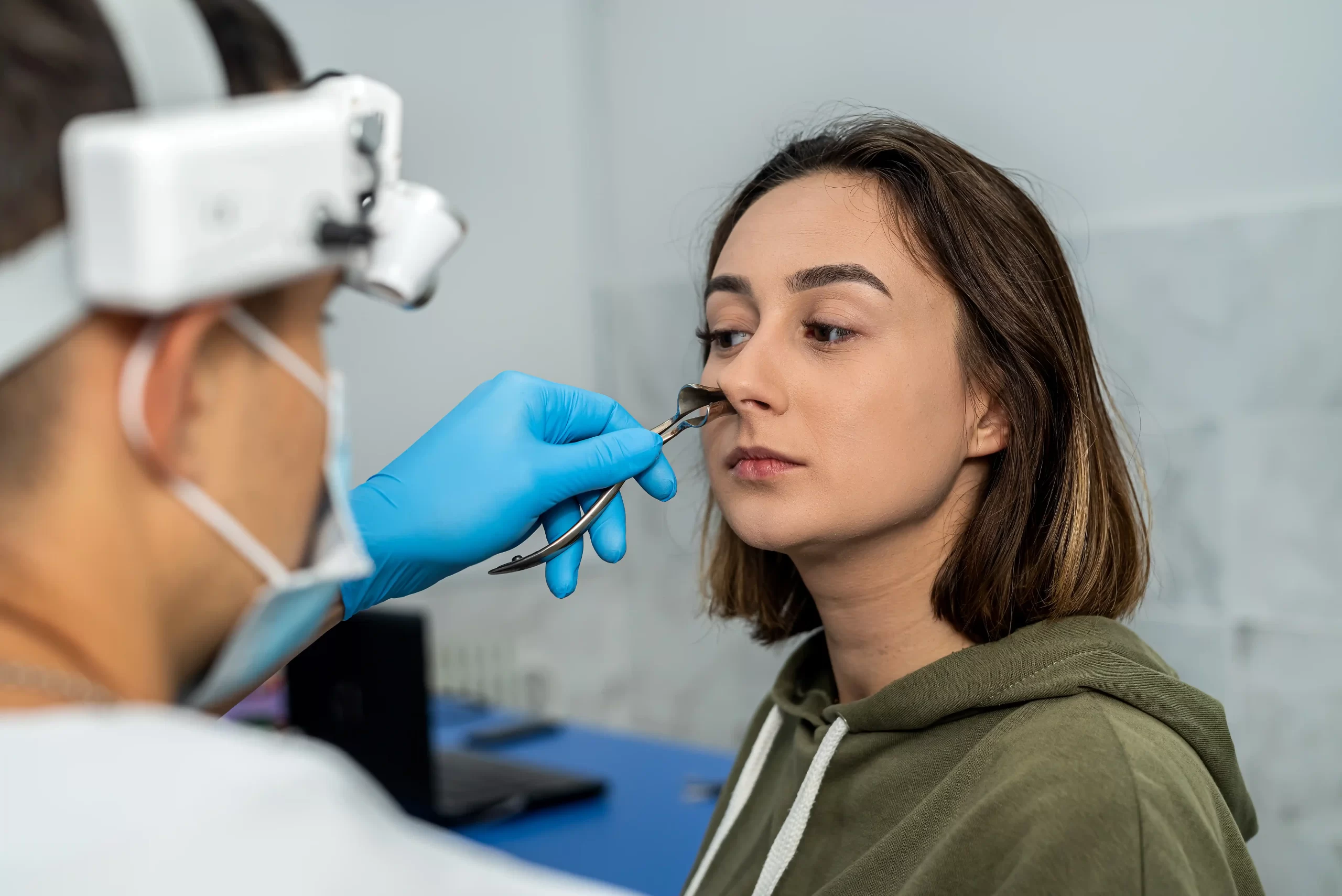
How is Orbital Surgery Done?
The Orbital Box Osteotomy is classified as a transcranial procedure. This designation is critical: it means the surgery cannot be performed solely from the “outside” of the face. It requires opening the skull (craniotomy) to access the roof of the orbits from above, necessitating the retraction of the frontal lobes of the brain. This interplay between neurosurgery and plastic surgery defines the complexity and risk profile of the operation.
Preoperative Planning and Virtual Simulation
In the contemporary era, the “eyeball” estimation method is obsolete. Every case undergoes high-resolution 3D CT imaging. The data is imported into Virtual Surgical Planning (VSP) software, where the surgeon performs a “mock surgery” on the computer screen.
The software allows the surgeon to define the exact cutting planes. The distance of the osteotomy from the orbital rim is critical—typically 15 to 17 mm—to ensure the cut is behind the globe’s equator but anterior to the optic canal, protecting the optic nerve. The surgeon calculates the precise volume of bone to be removed from the nasal midline (the nasion and ethmoid block). For example, to reduce an IOD from 45 mm to 25 mm, a 20 mm central block must be resected. The imaging identifies the position of the cribriform plate (where the olfactory nerves pass) and the height of the frontal sinus, allowing the team to plan the craniotomy to minimize dural tears and brain retraction injury.
Stage I: Access and Exposure
The procedure begins with the patient under general anesthesia, typically with neuro-monitoring to track brain activity. A zigzag or wavy incision is made across the scalp, from ear to ear. This design prevents a straight-line scar that would be visible through hair parting. The scalp flap is elevated (peeled forward) in the subgaleal plane to expose the frontal bone, the orbital rims, and the nasal bridge. The dissection continues laterally to expose the temporalis muscle and the zygomatic arch.
The neurosurgical team performs a bifrontal craniotomy. A section of the frontal bone (the “bone flap”) is removed and set aside. This window exposes the dura mater covering the frontal lobes. The dura is gently dissected from the anterior cranial fossa floor (which is also the roof of the orbits). Malleable retractors are used to lift the frontal lobes, exposing the orbital roofs from the inside of the skull. This step is crucial for making the superior osteotomy safely.
Stage II: The Osteotomies
The “Box” osteotomy gets its name from the four-wall cut that isolates the orbital cavity from the surrounding craniofacial skeleton.
Using a reciprocating saw or a piezoelectric bone cutter (which cuts bone but spares soft tissue), a transverse cut is made across the orbital roof. This cut connects the lateral and medial aspects of the orbit, strictly maintaining the pre-measured distance from the optic canal to avoid blinding the patient. The lateral osteotomy descends through the lateral orbital wall, separating the orbit from the sphenoid wing and zygoma.
Performed usually through the orbital rim, the inferior osteotomy traverses the floor of the orbit. Great care is taken to identify and protect the infraorbital nerve, which provides sensation to the cheek. If the nerve is in the path of the cut, it may be mobilized or, in rare cases, sacrificed if unavoidable (resulting in permanent numbness). The final medial osteotomy separates the medial orbital wall from the central nasal block.
Stage III: Midline Resection and Translocation
Once the orbits are osteotomized (cut), they are still held in place by soft tissue attachments. The surgeon then performs the central resection. A block of bone corresponding to the planned reduction (e.g., 20 mm) is removed from the central nasal area and the ethmoid sinuses. This creates the physical space for the eyes to move together. The two “orbital boxes” are physically mobilized and slid medially until they meet the new midline. This creates a “gap” on the lateral sides (the temples) where the orbits used to be.
Stage IV: Fixation and Reconstruction
Stability is paramount. If the bones shift post-operatively, the eyes will drift back (relapse). Titanium mini-plates and screws are used to bridge the osteotomy lines, locking the orbits into their new medial position.
The lateral gaps created by the movement must be filled to maintain structural continuity and temporal contour. Bone grafts are harvested from the craniotomy bone flap (split calvarial grafts) or, in children where the skull is thin, from the ribs. Moving the orbits medially often obliterates the nasal bridge. A cantilever bone graft is almost always required to reconstruct the nose and provide dorsal projection.
The medial canthal tendons, which anchor the inner corner of the eyelids, become lax when the bones are moved. They must be identified and re-anchored (transnasal canthopexy) with stainless steel wires to a central point to prevent telecanthus (soft tissue widening).
Stage V: Closure
The neurosurgeon checks the dura for any tears (CSF leaks) and repairs them watertight. The craniotomy bone flap is replaced and fixed. Drains are placed under the scalp to prevent hematoma, and the coronal incision is closed with sutures or staples.
Expertise and the Role of Dr. Burak Sercan Ercin
The Orbital Box Osteotomy is not a commodity service; it is a high-stakes intervention where the margin for error is measured in millimeters and the consequences of failure can be catastrophic (brain injury, blindness). Consequently, the choice of surgeon is the single most critical variable in the equation.
The Necessity of Microsurgical Proficiency
While OBO is primarily a bony surgery, the complications are often soft tissue in nature. A dural tear requires a watertight patch; a scalp wound breakdown requires a vascularized flap coverage. General plastic surgeons often lack the skill set for complex salvage.
Assoc. Prof. Dr. Burak Sercan Ercin distinguishes himself through a dedicated background in Reconstructive Microsurgery. His ability to perform free tissue transfer (connecting blood vessels under a microscope) is a vital safety net. If a complication arises—such as scalp necrosis or exposure of the hardware—a microsurgeon can harvest tissue from elsewhere in the body to cover the defect and save the patient from serious infection.
Dr. Ercin served as a Clinical Fellow at the Clinica Cavadas Reconstructive Surgery Unit in Spain. Dr. Pedro Cavadas is a global icon in reconstructive surgery, known for performing the world’s first double leg transplant and face transplants. Training in this environment exposes a surgeon to the absolute limits of human reconstruction. It implies a capability to handle “end-stage” deformities and intraoperative crises that would paralyze a less experienced surgeon. This pedigree is particularly relevant for OBO, where anatomical anomalies can be unpredictable.
Comprehensive Craniofacial Scope
Dr. Ercin’s practice at Medical Park Pendik is not limited to aesthetic rhinoplasty or liposuction. His clinical portfolio explicitly includes “Head and Facial Anomalies Treatment,” “Face and Jaw Surgery,” and “Maxillofacial Fixation”. He has documented success in treating hemifacial microsomia, a condition requiring asymmetric osteotomies and distraction osteogenesis. This demonstrates familiarity with the biological behavior of facial bone healing and the use of hardware in the pediatric and adolescent skeleton.
Dr. Ercin is also a member of the International Society of Orthoplastic Surgery. The principles of orthoplastics (combining orthopedic bone fixation with plastic soft tissue coverage) are the exact principles required for OBO. The “orbital box” is essentially an orthopedic manipulation of the skull, requiring plastic surgery coverage.
Risks, Complications, and Management Strategies
No discussion of Orbital Box Osteotomy is complete without a transparent analysis of risk. The “major adversity rate” for frontofacial surgery has been cited in literature as approximately 11%, referring to events requiring life-saving intervention or resulting in permanent sequelae.
Neurological Complications
A Cerebrospinal Fluid (CSF) Leak is the most common serious complication, occurring in up to 29.5% of cases in some series, though experienced centers report lower rates. It happens when the dura is torn during the separation of the orbit from the skull base. The olfactory rootlets (nerves of smell) anchor the dura to the cribriform plate. Moving the bone can shear these rootlets, creating a hole. Intraoperative repair is the first line of defense. Post-operatively, lumbar drains may be used to lower CSF pressure and allow the leak to seal. Persistent leaks may require re-operation.
A CSF leak creates a pathway for bacteria from the nasal cavity to enter the brain, leading to meningitis. Prophylactic antibiotics (e.g., Ceftriaxone, Vancomycin) are standard protocol. Pneumocephalus refers to air trapped inside the skull. While a small amount is normal post-craniotomy, “tension pneumocephalus” occurs if air builds up under pressure, compressing the brain. This is the primary reason for flight restrictions.
Ophthalmic Risks
The nerves controlling eye movement (III, IV, VI) travel through the superior orbital fissure, just behind the osteotomy cuts. Temporary stretching of these nerves is common, causing diplopia (double vision) or ptosis (droopy eyelid). Most palsies are neuropraxic (temporary stunning) and recover within 3-6 months. Permanent injury is rare but possible.
The risk of blindness is low (<1%) but non-zero. It can result from direct optic nerve trauma, or retrobulbar hematoma (bleeding behind the eye) that compresses the nerve. Immediate orbital decompression is the treatment for hematoma. The nasolacrimal duct (tear drainage system) can be damaged during the medial osteotomy, leading to chronic tearing (epiphora). This occurs in about 3.8% of cases and may require a secondary dacryocystorhinostomy (DCR) to create a new tear drain.
Aesthetic and Structural Complications
The soft tissues of the face have “memory.” If the canthal tendons are not securely anchored, the eyes can drift back toward a wide appearance (telecanthus), even if the bone stays in place. Implant infection can occur months or years later. Palpable hardware (screws you can feel under the skin) is a common complaint (92%) and may necessitate a minor surgery for removal once the bone is healed.
Recovery Trajectory: Timeline and Expectations
Recovery from OBO is a biphasic process: the acute physiological recovery and the chronic structural remodeling.
Phase 1: The Acute Phase (Weeks 1-2)
The patient is initially monitored in the ICU for 0-2 days. The head is elevated to 30 degrees to reduce intracranial pressure and swelling. Neurological checks are performed hourly. Swelling peaks massively around Day 3. The eyes are often swollen completely shut. This is expected and frightening for families, but resolves rapidly after day 4. Pericranial drains are removed once output decreases (usually Day 2-3). Once stable, the patient moves to a regular room. Ambulation (walking) is encouraged to prevent blood clots. Patients are typically discharged only when they are mobilizing well, eating a soft diet, and show no signs of CSF leak or infection, usually around Day 10-14.
Phase 2: The Consolidation Phase (Weeks 3-8)
A soft food diet is strictly enforced for 6 weeks. Chewing hard food transmits force through the maxilla to the orbits, potentially shifting the osteotomies. Patients must avoid heavy lifting or bending over. The hair can be washed gently. The scalp staples/sutures are typically removed around day 10-14.
Phase 3: Remodeling (Months 3-12)
The forehead will likely be numb initially. Sensation returns as the supraorbital nerves regenerate, often accompanied by “electric shock” sensations (dysesthesia). The coronal scar fades to a white line, hidden by hair. If plates are palpable or irritating, they are removed after 6-12 months.
The Economics of Craniofacial Surgery: A Global Comparison
For patients in the US and UK, the decision to travel to Turkey is often driven by the stark disparity in cost and access.
The Western Cost Prohibitive Model
In the United States, craniofacial surgery is among the most expensive medical undertakings. US hospitals bill separately for the surgeon, the assistant surgeon, the anesthesiologist, the facility fee (operating room time), and the ICU stay. Estimates typically place the surgeon fee between $20,000 and $40,000, anesthesia between $5,000 and $8,000, and the hospital/ICU stay between $80,000 and $120,000. The total often exceeds $120,000 to $170,000. While functional OBO is theoretically covered by insurance, high deductibles, co-pays, and the classification of certain “aesthetic” components (like canthopexy) can leave patients with massive out-of-pocket bills.
In the United Kingdom, the NHS covers OBO for severe syndromic cases (e.g., Crouzon, Apert). However, the waiting list for non-emergency craniofacial surgery can extend to 18-24 months due to capacity constraints. Private treatment in London (e.g., at Great Ormond Street’s private wing or The Portland) mirrors US prices, often exceeding £40,000 to £60,000.
The Turkish Value Proposition
Turkey has leveraged its lower labor costs and government incentives to create a high-volume, high-quality medical tourism sector. Turkish clinics typically quote a single “package” price. Dr. Ercin / Medical Park Pendik estimates for complex Craniofacial/Jaw Surgery generally range from $15,000 to $30,000. This typically includes the surgery, 1-2 nights ICU, 5-7 nights ward stay, nursing, medications, airport transfers, and sometimes hotel accommodation for the recovery period.
The primary savings come from the hospital facility fees and labor costs, not from using cheaper implants or technology. The titanium plates (e.g., Stryker, KLS Martin) and CT scanners (Siemens, GE) are the exact same global brands used in the US.
When we break down the costs, the surgical fee in the USA/UK private sector often starts at $30,000+, whereas it is included in the package in Turkey. Hospital/ICU costs in the West are itemized and can exceed $80,000, but are included in the Turkish package. Pre-op imaging can cost thousands in the West but is often included or very low cost (around $300) in Turkey. The total cost comparison shows a range of $120,000 – $170,000 in the West versus $15,000 – $30,000 in Turkey, with wait times reduced from months/years to just weeks.

Logistical Protocol for International Patients
Traveling for major surgery requires military-grade planning. The following protocol outlines the necessary steps for a patient traveling from the UK or USA to Dr. Ercin.
Step 1: Remote Assessment
Patients must obtain their DICOM files (CT scans) from their local provider and upload them to Dr. Ercin’s team. A video consultation determines candidacy, where Dr. Ercin evaluates the soft tissue laxity and discusses the risks. A fixed quote is provided. Payment is typically made at the clinic, often with a deposit to secure the date.
Step 2: Arrival and Pre-Op (Days -2 to 0)
Istanbul has two major airports (IST and SAW). Medical Park Pendik is located on the Asian side, closer to Sabiha Gokcen Airport (SAW). The hospital usually arranges a driver to pick the patient up from the airport. Blood tests (hemogram, coagulation profile), infectious disease markers, and cardiac consultation (ECG/Echo) are performed. Anesthesiology clearance is final.
Step 3: Surgery and Hospitalization (Days 1-14)
It is strongly advised to travel with a support person. The hospital rooms are typically private and include a bed for a companion. English-speaking coordinators or translators are assigned to international patients to facilitate communication with nursing staff.
Step 4: The “Fit to Fly” Critical Window
This is the most misunderstood aspect of craniofacial medical tourism. Patients cannot fly home immediately after hospital discharge. During craniotomy, air enters the skull (intracranial air). At sea level, this is harmless and absorbs over time. However, airplane cabins are pressurized to equivalent altitudes of 6,000-8,000 feet. According to Boyle’s Law, gas expands as pressure drops. Trapped intracranial air will expand by approximately 30% at cruising altitude. This expanding air inside a closed skull acts like a growing tumor (Tension Pneumocephalus), compressing the brain and potentially causing herniation, seizures, or death.
The UK Civil Aviation Authority (CAA) recommends a minimum of 7 days post-craniotomy before flying. Neurosurgical consensus often advises waiting 2 to 4 weeks depending on the amount of air seen on the post-op CT. Patients must plan to stay in a hotel in Istanbul for at least 7-10 days after leaving the hospital. A control CT scan is often required to confirm the air has resorbed before the surgeon issues a “Fit to Fly” certificate. A total trip duration of 21-28 days is recommended (2 days pre-op + 14 days hospital + 7-12 days hotel recovery).
The “Bella Eyes” Trend vs. Medical Necessity
A growing segment of Dr. Ercin’s inquiries comes from patients seeking OBO for purely aesthetic reasons—specifically to achieve the “wide-set” or “alien” look popularized by social media filters (often termed “Bella Eyes”).
The Ethics of Aesthetic Osteotomy
True OBO involves significant risk to life and vision. Performing a craniotomy solely for aesthetic enhancement in a patient with normal orbital anatomy is highly controversial and generally discouraged by the international surgical community. Patients with hypotelorism (eyes too close together) may be candidates if the condition causes functional issues or severe aesthetic disharmony. However, for patients with normal IOD who simply want an “exotic” look, OBO is too aggressive. The risk-benefit ratio does not justify the potential for brain infection or blindness.
The Soft Tissue Alternative
Dr. Ercin offers less invasive alternatives for this demographic. Canthoplasty or “Fox Eyes” procedures surgically alter the lateral canthal tendon (the outer corner of the eye) and lift the brow. This changes the apparent shape of the eye, elongating it and tilting it upward to mimic the look of wide-set eyes without breaking bone. Strategic fat grafting to the lateral cheekbones can also create the illusion of facial width. Patients must clearly understand the distinction: OBO is reconstructive skull surgery; “Fox Eyes” is aesthetic soft tissue surgery. Dr. Ercin’s expertise allows him to guide patients toward the safer, appropriate option.
Conclusion
The Orbital Box Osteotomy stands as a testament to the capabilities of modern surgery—the ability to safely dismantle and reconstruct the human face to restore essential sensory function and psychosocial well-being. For patients suffering from hypertelorism, the procedure is life-changing, offering a release from functional visual deficits and the stigma of facial deformity.
However, the barriers to this care—financial and logistical—are formidable. The emergence of high-quality, accredited centers in Turkey offers a solution. Dr. Burak Sercan Ercin, with his distinguished background in microsurgery and complex reconstruction, and the institutional backing of Medical Park Pendik, represents a viable and high-standard option for international patients. By combining the rigor of European-standard microsurgery with the accessibility of the Turkish medical tourism model, Dr. Ercin’s practice bridges the gap between need and access.
For the international patient, the journey requires diligence. It is not a simple “medical holiday” but a major surgical undertaking requiring weeks of commitment, strict adherence to flight safety protocols, and a comprehensive understanding of the risks. With the right team and the right preparation, the Orbital Box Osteotomy offers a horizon of new possibilities for those with complex craniofacial anomalies.
To summarize the logistics, choosing Turkey for this procedure involves comparing a specialist in Craniofacial Plastic Surgery (USA/UK) with a Reconstructive Microsurgery & Craniofacial specialist (Dr. Ercin). Accreditation standards are comparable (JCI vs State Licensing), but costs drop dramatically from $120,000+ to $15,000 – $30,000. Inclusions in Turkey cover VIP transfers and accommodation support, unlike the surgery-only fees in the West. Wait times are weeks rather than months or years, though the minimum stay is longer (21-28 days) due to flight safety.
Regarding risks, CSF Leak occurs in 5-30% of cases and is managed via intraoperative repair or drains. Infection rates are 5-10%, managed with antibiotics. Diplopia is common but usually transient. Blindness is rare (<1%) but possible, mitigated by precise osteotomy design. Relapse is variable and prevented by rigid fixation.
This content was written by Assoc. Prof. Dr. Burak Sercan Erçin in line with clinical experience and current medical literature. It is intended for general informational purposes only and does not constitute medical advice. A personal consultation with Dr. Erçin is recommended for individual assessment.
Faculty Member · Bahçeşehir University
Graduate of Ege University Faculty of Medicine, Assoc. Prof. Dr. Erçin completed advanced fellowships at Tampa General Hospital (USA) under Dr. Deniz Dayıcıoğlu in breast reconstruction and burn surgery, and at the clinic of Dr. Pedro Cavadas in Valencia, Spain in reconstructive microsurgery. After passing the EBOPRAS examination in 2018, he joined Bahçeşehir University as a faculty member and continues his private practice on Bağdat Avenue, Istanbul, specialising in face, breast and body aesthetics alongside complex reconstructive surgery.